 Notes: nsb neuroscience - swallowing & speech
Similar resources:
Y2, Y2Notes, Y2 NSB, Neuro Notes, Neuroscience, Christopher Yeo, Chris Yeo, NSBMovement, NSBNeuro
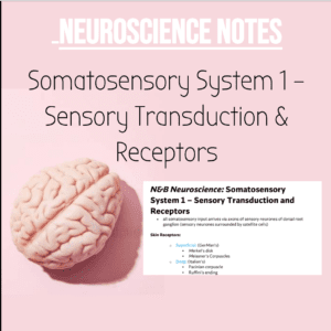
N&B Neuroscience: Swallowing and Speech
Swallowing:
https://www.youtube.com/watch?v=YQm5RCz9Pxc
1. Oral Phase
o Jaw needs to open (to eat food)
▪ lateral pterygoid (CN Viii)
▪ digastric anterior belly (CN Viii)
▪ mylohyoid (CN Viii)
▪ geniohyoid (C1 cervical plexus)
o Jaw needs to close (to eat food)
▪ masseter (CN Viii)
▪ temporalis (CN Viii)
▪ medial pterygoid (CN Viii)
o Salivation to break down food into a bolus
o Intrinsic and Extrinsic muscles of the tongue contract to make the
tongue a downward slope for the food to pass to the oropharynx
▪ intrinsic muscle (CN 12) – superior + inferior longitudinal +
transverse muscles
▪ extrinsic muscles (CN 12) – styloglossus, genioglossus
o bolus touches palatoglossal arch (now in pharyngeal phase)
2. Pharyngeal Phase
o When bolus activates CN 9 it then activates CN 10.
▪ Nasopharynx action:
▪ vagus nerve stimulates uvulae to contract up, blocking the
nasal pharynx.
▪ vagus contracts levator veli palateni, pulling up the soft
palate
▪ trigeminal nerve (Viii) contract tensor veli palateni, so soft
palate is elevated even more.
▪ Larynx action for trachea:
▪ adduction of vocal cords (cricothyroid, aryepiglottic)
▪ epiglottis folds down (elastic cartilage)
▪ aryepiglottic muscles and cricothyroid muscles help to
close larynx.
▪ Pharynx:
▪ Outer longitudinal layer (elevate pharynx and larynx)
▪ stylopharyngeus (CN 9)
▪ salpingopharyngeus (CN 10)
▪ palatopharyngeus (CN 10)
▪ Inner Circular Layer (peristalsis of bolus)
▪ superior pharyngeal constrictors
▪ middle pharyngeal constrictors
▪ inferior pharyngeal constrictors
▪ larynx moved up and anteriorly by suprahyoid muscles
N&B Neuroscience: Swallowing and Speech
▪ cricopharyngeus (upper oesophagus sphincter) (CN 10)
3. Oesophageal Phase
o primary peristalsis is a continuation of pharyngeal peristalsis
o lower oesophageal sphincter relaxes
o bolus enters stomach
o secondary peristalsis (local reflex) when bolus is stuck in oesophagus.
• NTS and trigeminal nuclei receive information from the tongue and pharynx
and larynx, and the nucleus ambiguus and hypoglossal nuclei supply the
muscles in those structures
• swallowing central pattern generator is formed by reticular formation neurones
• brainstem strokes often produce dysphagia
• swallowing area in brain is bilateral, but one hemisphere is preferentially
activated during swallowing. (just behind Broca’s area and extends into insular
cortex)
• left sided cortical strokes often produce dysphagia but can recover over a few
weeks as other side of brain works.
• aphagia- loss of swallowing and eating
Speech:
• speech is producing elemental sounds (phonemes) that combine to make
meaningful sounds (morphemes) and sequences of morphemes make up
words.
• speech requires accurate control of:
o respiration- breathing control in speech
o phonation- sound production in the larynx
o resonance- selective amplification of some sounds
o prosody- varying intonations, stress, and rhythm
o articulation- movement timing in speech structures
▪ disruption of any of these produce dysarthria (poor generation of
phonemes) (can have flaccid or spastic dysarthria (UMN vs
LMN))
• Dysarthria: (loss of motor control of speech)
o Flaccid Dysarthria- weak control of speech musculature (LMN lesion)
o Spastic Dysarthria- tension in muscles (UMN lesion)
• dysphasia or aphasia are losses of speech and language from higher aspects
• Broca’s Area:
o left inferior frontal gyrus
o expressive or motor speech area
o patients understand speech, but can’t express speech (can’t find the
words)
▪ Severe Expressive Aphasia (Broca’s or Motor aphasia) (tono
tono)
▪ Expressive Aphasia (Broca’s or Motor aphasia) (can’t find the
words)
N&B Neuroscience: Swallowing and Speech
• Wernicke’s Area:
o comprehension and meaningful speech
▪ Receptive Aphasia (Wernicke’s or Fluent aphasia) (fluent speech
but doesn’t make any sense)
• Wada Test- proves that language is left side of the brain (sodium amytal
anaesthetises one hemisphere of the brain)
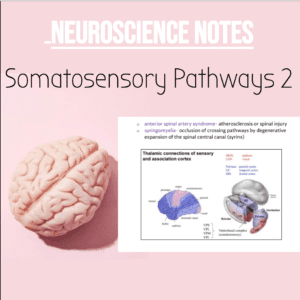
• Wernicke-Geschwind Model of Speech:
o Posterior Speech Area (angular gyrus, temporal and parietal regions)
o auditory and visual information converge here.
o arcuate fasciculus
o extreme capsule
o reading and speaking- angular gyrus (visual and auditory information)
o hearing and speaking- angular gyrus (visual and auditory information)
4 Types of Aphasia: