 Notes: nsb neuroanatomy brainstem
Similar resources:
- All
- CPSA
- Lecture Notes
- Videos
NSB Neuroanat Notes: Forebrain 2
NSB Neuroanat Notes: Forebrain 3
NSB Neuroanat Notes: Introduction
Video: Cranial Nerves | NSB

Neuroanatomy Summary
Y2 OCaPE: NSB 5 Head & Neck Anatomy
Video: Circle of Willis Summary | NSB
NSB Neuroanat Notes: Brain & Spinal Cord
NSB: Cerebrum
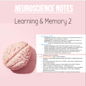
NSB Neuroscience Notes: Learning & Memory 2
Video: Cerebellum Overview | NSB
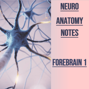
NSB Neuroanat Notes: Forebrain 1
Y2, Y2Notes, Y2 NSB, Y2NeuroAnat neuroanatomy Patrick anderson
Somatosensory Systems: (ascending tracts)
• anterior spinal artery supplies the ventral grey horns, lateral corticospinal tract
o if the anterior spinal artery is blocked (spinal stroke), the great
descending motor tracts and main ascending pain pathways will all die
• posterior intercostal arteries → radicular arteries → anterior spinal artery
o Artery of Adamkiewicz (largest radicular artery – T10 level)
1. Primary Afferent Fibres (Sensory Neurones): (have cell bodies in sensory
ganglia)
• 1a afferent- ventral horn, largest and fastest axon, myelinated, proprioception
• A beta afferent- dorsal horn, detects touch, myelinated
• A delta afferent- thin myelin, detect initial pain
• c-fibre afferent- thinnest diameter and slowest, unmyelinated, nociception
2. Second Order Neurones:
• are on the same side of the CNS and their axons cross the midline to reach the
thalamus
3. Thalamic Neurones:
• send information from the thalamus to the somatosensory cortex for us to feel
Spinothalamic Tract:
• nociception (temperature, touch)
1. c-fibres and A delta fibres detect pain from skin or muscle
2. synapse in superficial dorsal horn (laminae 1+2)
3. decussation occurs where the primary afferent fibre enters the spinal
cord
4. secondary order neurone goes up the spinothalamic tract
5. collaterals from the second order neurone go to brainstem reticular
formation (keeps you conscious)
6. second order neurone synapses in the thalamus
7. thalamic neurone goes to the somatosensory cortex
hypoglossal nucleus is for the hypoglossal nerve (12), which supplies the
muscles of the tongue
o if there is a lesion on the neurone, the tongue points to the side of the
lesion
o all somatic motor nuclei are near the midline
• Nucleus ambiguus- cranial nerves, 9, 10, 11 and supplies the muscles of the
pharynx, larynx, and soft palate (speech and swallowing)
Pupillary Light Reflex:
1. shine light in left eye
2. ganglion cell in retina in left eye carries AP down the optic nerve to pretectal
nucleus
3. pretectal nucleus has two nerves from it, one ipsilateral and one contralateral.
1. ipsilateral = to Edinger-Westphal nucleus to oculomotor nerve in left
eye to ciliary ganglion to constrictor pupillae muscle, causing direct
response of left eye constriction
2. contralateral = to Edinger-Westphal nucleus to oculomotor nerve in
right eye to ciliary ganglion, causing indirect response of right eye
constricting
• ipsilateral oculomotor palsy- double vision, down and out, enlarged pupil, ptosis
Neuroanatomy: Brainstem 2
Cerebellum:
• coordinates movement (via thalamus and cerebral cortex)
• Floccular-nodular Lobe:
o vestibulocerebellum (keeps balance)
o truncal ataxia
o nystagmus
o eye movement problems
• Medial Longitudinal fasciculus- connects nuclei of 3,4,6,8 (vestibulo-ocular
reflexes)
• Deep Nuclei of the Cerebellum:
o Dentate (projects to thalamus)
o Emboliform
o Globose
o Fastigial (projects to vestibular nuclei and reticular formation)
• Layers of Cerebellar Cortex:
o Molecular layers
o Purkinje Cells
o Granular Cells Layer
Neuroanatomy: Brainstem 2
Input to cerebellum comes from:
o climbing fibres- inferior olivary nucleus
▪ olivocerebellar
▪ through inferior cerebellar peduncles
▪ each purkinje cell has one climbing fibre (these are excitatory and
convey information about motor errors)
o mossy fibres- everywhere else (pons, medulla, etc.)
▪ spinocerebellar fibres- locomotion
▪ pontocerebellar fibres- non-stereotyped movement (from
pontine nucleus through middle cerebellar peduncles and become
mossy fibres in contralateral cerebellum)
▪ cerebral cortex → pontine nuclei → cerebellum (cortico-ponto-
cerebellar pathway)
Spinocerebellar Tract:
o Dorsal Spinocerebellar Tract
▪ C8-L2 (Clarke’s Column)
▪ proprioceptor signals of upper limbs enter spinal cord via
primary afferent neurone
▪ second order neurone goes to dorsal SCT on same side
▪ second order neurone goes through the inferior cerebellar
peduncle to cerebellar cortex
o Ventral Spinocerebellar Tract
▪ below L3
▪ proprioceptor signals enter spinal cord via primary afferent
neurone
▪ second order neurone enters ventral spinocerebellar tract
on contralateral side
▪ neurone in ventral SCT enters through superior cerebellar
peduncle
▪ neurone crosses behind brainstem to go to cerebellar
cortex on other side
o Cuneocerebellar Tract
▪ above C8
▪ proprioceptor signals enter spinal cord via primary afferent
neurone
▪ primary afferent goes to fasciculus cuneatus then
synapses with accessory cuneatus nucleus
▪ second order neurone goes to inferior cerebellar peduncle
and goes to cerebellar cortex
Neuroanatomy: Brainstem 2
• Cerebellar hemisphere lesions:
o lateral part- intention tremor, dysdiadochokinesia (can’t perform rapid
muscle movements – flipping hand over each side), speech problems
o anterior lobe- gait ataxia (unsteady walking- alcoholics)
• Vestibulospinal Tract:
o keeps you upright
o not involved in voluntary movement
o 4 neurones in descending tract (ipsilateral)
1. input from flocculo-nodular lobe of cerebellum
2. neurone synapse in lateral vestibular nucleus
3. axon in vestibulospinal tract
4. interneuron in spinal cord
5. spinal motor neurone to erector spinae muscle (head and neck
muscles to keep you upright)